The ICA MRA includes original segment labels. The DSA examples are unannotated reference images; the reading prompts are teaching guidance, not verified pixel annotations.
AN INTERACTIVE GUIDE TO CEREBRAL ANGIOGRAPHY
Understand the projection.
One patient's vascular tree, from real CTA. Move the C-arm and watch anatomy become an angiogram.
PATIENT-MATCHED CTA 02 / ARTERIAL & VENOUS
01 3D anatomy
PATIENT AXES
Drag to orbit · Scroll to zoom · Click a vessel
ICA / MCAACA / communicatingPCA / vertebrobasilarCerebellarVeins
02 Angiographic projection
AP · 0°
RLSUBTRACTED
LEFT CAROTID TREE · SEGMENTS & ORIGINS
Middle cerebral artery
About this model · Patient-matched CTA.
FROM THE MODEL TO THE IMAGE
Real angiography. Real variation.
The CTA MIP · real mode in the projection panel already shows this patient's actual CTA, projected at the current C-arm angle and restricted to the intracranial space. Below, compare with published catheter angiograms. These are independent reference cases, not images of the CTA patients, and their acquisition angles are not calibrated to the simulator.
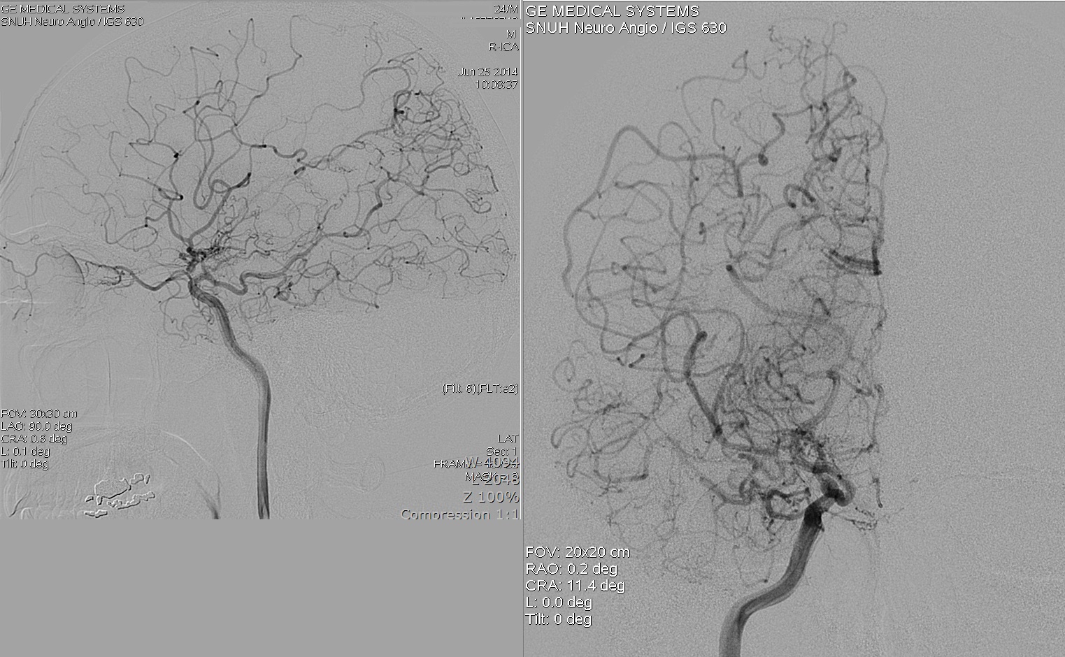
Image delivery is unavailable. Open the original teaching image using the source link below.
PATIENT-MATCHED ANATOMY
Every view comes from one real scan.
For each case, the vessels, skull, slices and the CTA projections derive from the same TopCoW/TopBrain CTA, in one coordinate frame, and the brain is a detailed template fitted to that patient's brain. Named segments are the TopBrain expert labels. Branches beyond those labels are traced from the CTA's own contrast and assigned to their labelled parent.
TopBrain release ↗ · TopCoW scans & masks ↗ · Centreline graphs ↗HOW TO USE LEARN CEREBRAL
From 3D anatomy to the angiogram, in four panels.
Learn Cerebral shows one patient's brain vessels three ways at once: as 3D anatomy, as the angiogram a C-arm would record, and as the C-arm itself positioned around the patient. Change the angle in any of them and the others follow.
About a minute. Use the arrow keys to step through, Esc to leave.
- 1
Choose a patient and an injection
Pick a CTA case from the bar above the panels. Then choose which tree fills with contrast in panel 02: Left ICA, Right ICA, Vertebral, All arteries or the Venous phase.
- 2
Explore the 3D anatomy (01)
Drag to rotate, scroll or use + / − to zoom, and click a vessel to select it; it turns lime in every view. Use the checkboxes to add the brain, skull or veins, and the Labels menu to show all labels, only the highlighted vessel, or none. More options, including CTA slices and brain opacity, are under Brain, CTA slices & centrelines.
- 3
Set the C-arm angle (03 and 04)
Move the RAO / LAO and Cranial / caudal sliders, click a standard view, or click anywhere on the angle map. In the Table & C-arm panel you can also drag the green detector directly; drag elsewhere to look around the room. ▷ Tour plays through the standard views.
- 4
Read the angiogram (02)
The projection is computed from the patient's own scan at the current angle. Subtracted shows contrast only, like DSA; Unsubtracted keeps the skull; CTA MIP projects the real CTA. Click a vessel in the image to identify it. Color tints each territory and the field size zooms the detector.
- 5
Lock the views together
Press 🔓 Lock 01 · 02 · 04 in the anatomy panel. Dragging the 3D anatomy or the angiogram then turns the C-arm, so the anatomy is always seen from the detector, exactly as in the image.
- 6
Learn the segments
Under the angiogram, Segments & origins lists every part of the selected tree, its common names (for example C2 petrous or M1 sphenoidal) and the branches it gives rise to. Vessel spotlight describes whichever vessel is selected.
- 7
Test yourself
Name the vessel highlights a random vessel inside a pulsing ring. Rotate and re-angle to trace it back to its origin, then choose its name. Real imaging compares the model with published angiograms, and the Field guide summarizes the key ideas.
Words you will see
- AP
- Anteroposterior: the beam passes front to back, giving a frontal view.
- RAO / LAO
- Right / left anterior oblique: the C-arm turned so the detector sits toward the patient's right or left. LAO 90° is a lateral.
- Cranial / caudal
- The detector tilted toward the head or the feet.
- Subtracted (DSA)
- An image showing only contrast-filled vessels, with bone removed.
- MIP
- Maximum-intensity projection: the brightest CT value along each ray.
- Foreshortening
- A vessel running toward the beam looks shorter than it is. Changing the angle reveals its true length.
- Isocentre
- The point the C-arm rotates around, here at the Circle of Willis.
Keyboard
- ← → ↑ ↓
- Rotate the 3D anatomy, or change the angle when the angiogram, angle map or C-arm is focused (hold Shift for larger steps).
- + −
- Zoom the 3D anatomy.
- Tab
- Move between panels and controls.
- Esc
- Leave the guided tour.
Tips
- Start in AP with one carotid injection, then add 20–30° of obliquity and watch overlapping branches separate.
- Turn on Beam in the anatomy panel to see the X-ray path through the head.
- Use Full screen on smaller laptops.
FIELD GUIDE
Read the anatomy before the image.
Name the detector side
LAO and RAO name the side of the patient nearest the detector. Cranial and caudal name where the detector is tilted. In the Table & C-arm panel the patient and table stay fixed; only the arm rotates about the isocentre. The projection is displayed as seen from the detector, so in AP the patient's right is on your left.
Depth becomes overlap
A projection collapses three-dimensional anatomy onto a plane, magnified by the beam's divergence from the source. AP separates left from right; lateral separates anterior from posterior. Oblique and angulated views open branch origins hidden by overlap and foreshortening.
Subtracted, unsubtracted, real
Subtracted shows contrast path length through the segmented vessels, like a road-map of DSA. Unsubtracted adds a radiograph computed through the real CTA, so the skull base and calvarium overlie the vessels. CTA MIP is the patient's actual CTA, projected at the same angle.
Choose an injection
Compare each internal carotid tree with the vertebrobasilar tree, then show all arteries to inspect the communicating connections. The venous option shows labelled sinuses, deep veins and CTA-traced cortical veins. Filters select anatomy; they do not model flow, collateral filling or timing.
Where labels end
Expert labels stop at M3, A3 and P3–P4. Beyond them, branches visible on the CTA are traced and grouped by parent: “MCA cortical branches”, “ACA distal branches” and so on. Their completeness depends on the scan's contrast. Case 002 shows many; case 008 shows fewer.
Brain and skull
The cortex, cerebellum and brainstem are a detailed brain template fitted to each patient's own brain, so the gyri sit where that patient's brain is; the skull comes straight from the CTA. Choose a skull cutaway and a see-through or solid brain under “Brain, CTA slices & centrelines”, then use “Look along imaging axis” to see what the beam passes through.
ABOUT & REFERENCES
Real anatomy, built for neurovascular learning.
Learn Cerebral is an independent educational project that pairs a patient's 3D cerebral vessels with the angiogram a C-arm would produce from any angle. It is inspired by the anatomy–projection–C-arm teaching approach of LearnCoronary; all code, models and imagery here are original or openly licensed.
Created by Sam Bogdanov · Built with Claude Opus 5.5 and GPT Sol-6.1 / Astra-6
How the models are built
Cases: six TopCoW CTA scans (002, 003, 008, 012, 021, 022) from the TopBrain release, chosen to show common variants. Vessels: per-label surfaces of the TopBrain v2 (36-class) arterial and v1 venous expert annotations, extracted at native resolution. Distal branches: CTA voxels above a case-specific threshold, connected to the labelled tree and assigned to the nearest labelled parent by geodesic propagation. Dural sinuses: venous channels grown from the labelled sinuses and named (torcula, transverse and sigmoid sinuses) by position and calibre after registration to standard space. Brain: the ICBM152 2009a template cortex, cerebellum and brainstem, fitted to each patient's brain with an affine and then a symmetric diffeomorphic (SyN) registration; ventricles from the patient's CSF on CTA; deep grey nuclei from the warped Harvard–Oxford atlas. Skull: surfaced from the native-resolution CTA, limited to the scan's field of view. Projections: perspective C-arm geometry (SID 1100 mm, SOD 750 mm), Beer–Lambert path length through vessel surfaces, and ray-marched DRR/MIP through the 1 mm CTA.
About the simulation
Every view is generated from real CT angiography, so vessel courses, overlap and foreshortening reflect a real patient rather than an idealized drawing. The projection is a teaching simulation designed to build intuition for how acquisition angles shape the image: it shows where contrast-filled vessels project, not the full physics of an X-ray system, blood flow or contrast timing. Distal branches are traced directly from the scan's contrast, and preset angles are common starting points for exploring each circulation. Learn Cerebral is intended for education, not for clinical decision-making.
What's next
Planned additions include review of the anatomy and segment naming with neurointerventional colleagues, more patient cases from the TopBrain collection, including MRA, timed arterial-to-venous contrast phases, more physically realistic image contrast for bone and iodine, and vascular territory maps on the fitted brain.
Data sources & licences
- CTA scans and TopCoW Circle of Willis masks: Yang et al., TopCoW 2024 MICCAI Challenge Data Release, University Hospital Zurich / University of Zurich. Dataset · Terms. Open use with source attribution; commercial use requires data-owner permission.
- Whole-brain vessel labels: TopBrain Challenge Data Release (Yang K, Shi P, Huang H, et al. TopBrain segmentation challenge for whole brain vessel anatomy. medRxiv 2026). Dataset · Terms. Same terms as TopCoW.
- Centreline graphs: Musio et al., Circle of Willis Centerline Graphs and Morphometric Features. Dataset, CC BY-NC as supplied. Supplied licence.
- Harvard–Oxford subcortical atlas (FSL; via the neurodata/neuroparc collection), FSL licence, non-commercial. Diedrichsen 2009 probabilistic cerebellar atlas (via DiedrichsenLab/cerebellar_atlases), CC BY. See atlas sources.
- ICBM 2009a nonlinear symmetric brain template (Fonov et al., NeuroImage 2009/2011), Copyright © 1993–2004 Louis Collins, McConnell Brain Imaging Centre, Montreal Neurological Institute, McGill University; used under its permissive licence (distributed with nilearn). Registration with DIPY (BSD).
- Rendering uses Three.js 0.180.0 (MIT).
Reading and anatomy references
New here? Take the one-minute guided tour.